

Capnography - See it for Free
| This page is from our TMC course |
Capnography isn't as heavily tested as it should be. A noninvasive tool for monitoring CO2 (ventilation status) ... as powerful a measure as that other noninvasive vital sign (pulse oximetry).
See Also: Capnography (Equipment) for details about types of capnography (sidestream, mainstream) and terms you should know (quantitative vs qualitative, colorimetric, etc.)
|
Normal Values: 35-45 mmHg (no different than PaCO2) |
Uses
- Monitoring patients during procedures requiring sedation
- When patients are given medications that can suppress the respiratory drive, a specialized nasal cannula is used to monitor adequate ventilation
- When patients are given medications that can suppress the respiratory drive, a specialized nasal cannula is used to monitor adequate ventilation
- Cardiac arrest
- Monitoring tracheal tube placement
- Monitoring CPR quality
- < 10 mmHg, consider improving CPR quality
- Can detect the return of spontaneous circulation (ROSC)
- A quick increase and sustaining in CO2 value is a strong indicator of ROSC
- A sharp decrease in CO2 may indicate a loss of ROSC
- Mechanically ventilated patients
- Real-time monitoring of patient's ventilatory status
- Helpful for noninvasive trending of CO2 throughout the day
- Verify endotracheal tube placement using disposable colorimetric
capnometry - A specialized adaptor is attached to the endotracheal tube after intubation which can detect the presence of CO2
- When CO2
is detected it causes the device to indicate a color - It can take several breaths, especially after cardiac arrest, for the color to sufficiently change
- Use the following to help you remember (note: technically the color is closer to "tan" when CO2 is inconclusive, 3-15 mmHg):
Color is Purple
(ETCO2 < 3 mmHg)Purple is Problem
Endotracheal/Tracheal tube is not in the trachea
- Recommend removal of tube, reintubation
- If trach, likely in false tract (tissue) or embedded in posterior wall of trachea. Remove trach, recommend speciality trach
Color is Yellow
(ETCO2 > 15 mmHg)Yellow is Yes
Endotracheal/Tracheal tube is likely in the trachea
- Recommend further confirmation, including chest radiograph to verify position
- Recommend removal of tube, reintubation
Interpreting Values
Decreased ETCO2
- Cardiac arrest!
- Pulmonary Embolism
- Hyperventilation
- Bronchospasm
- Mucus Plugging
Increased ETCO2
- Respiratory insufficiency (shallow breathing: hypoventilation)
- Respiratory depression (insufficient RR, especially during procedural sedation, overmedication)
- Fever, agitation, and pain lead to an increased production of carbon dioxide causing an increased ETCO2
Interpreting Graphics
The NBRC test could certainly have some basic waveform capnography on it. We recommend reviewing the main elements of common scenarios:

Unknown source
A few things to note here:
- It is not unusual to be provided a view of the real-time capnogram (left-side) alongside the trend (right-side). Be sure to look at both. Sudden changes and Gradual changes are both important.
- Look at the letter D in the diagram - remember that this is the reading of end-tidal CO2 (end of tidal volume - CO2), usually the highest peak)

You can recognize hypervention as the plateau gets lower and lower, indicating a decreasing CO2 (the patient is blowing off CO2)
If you are given a trend, as you see in this image, you see that the CO2 has decreased steadily.

As you might expect, this is the opposite of hyperventilation. In hypoventilation, the plateau gets higher and higher, indicating a worsening fo respiratory status (hypoventilation)
This is a common capnogram for testing purposes.
|
Example scenario: The Respiratory Therapist is monitoring a patient who has received multiple doses of midazolam (Versed). The following capnogram is noted:
The Respiratory Therapist should: A. begin compressions This type of question would just be looking for you to recognize this as hypoventilation during procedural sedation. While we don't know a RR and have no other information in this case, the only answer option that makes sense is C. |
Source: derangedphysiology.com
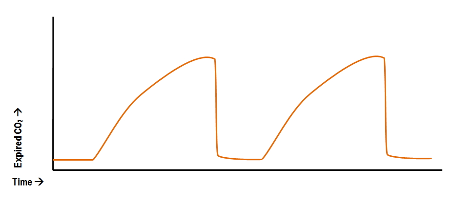
This image is classic shark-fin (or sawtooth) appearance, common with bronchospasm/bronchoconstriction. This happens due to a more turbulent mixing of deadspace and alveolar air, "smoothing" the rise in CO2 (normal is a more sudden rise)

A sudden drop to zero should have you thinking, "Oh, no! Something's not good."
Considerations include:
- Sudden ventilator disconnect
- Airway out of place (decannulation, extubation)
- Loss of pulse (start compressions!)
- It's also possible with severe, life-threatening air-trapping

During compressions, a sudden increase in PaCO2 may indicate a return of spontaneous circulation (ROSC) which is detectable before the pulse is rechecked. Note that this sudden increase will be less apparent if high-quality compressions with effective bag-mask (or artificial airway) ventilation is occurring.

Source: derangedphysiology.com
Note: dip/scoop in waveform, called a curare cleft
Action: patient is attempting a breath. Ensure synchrony or if undesired may need to further sedate

