

ECMO for ACCS
VV-ECMO, VA-ECMO, ECCO2R
|
Oakes Academy Tip ECMO is fair game on the ACCS. The updated ACCS places greater emphasis on ECMO, but don't overthink it. Focus on core concepts. When to start it? Which type makes sense? What are the key variables to change and how (flow vs sweep vs vent) in response to an ABG? What are some key troubleshooting issues and steps?
|
VV (Venovenous) or VA (venoarterial)?
Something to know: the letter indicates drainage (1st letter) and return (2nd letter)
Ex: VV-ECMO is flow that leaves from a vein, then returns to a vein, both on the right side of the heart.
| VV ECMO | VA ECMO | |
| Purpose | LUNG Support | HEART + LUNG Support |
| Blood leaves from | Vein | Vein |
| Blood returns to | Vein | Artery |
| Examples: |
Severe ARDS Pulmonary injury/trauma |
Cardiogenic shock |
The WHEN of ECMO
Our thought is this: try all the things and when they don't work, try ECMO. The ACCS seldom jumps quickly to ECMO - optimize everything - but then ECMO should be a valid option to consider.
Want an exception? A witness cardiac arrest, especially if in-hospital. Recommend cannulation, avoid if a "red flag" exists.
Classic example:
Severe ARDS + refractory hypoxemia (hypercapnia to a lesser degree) despite optimized conventional therapy → consider VV ECMO.
Other situations may include severe potentially reversible respiratory failure such as:
- Severe ARDS (common on test!)
- Severe asthma (common on test!)
- Refractory hypoxemia or hypercapnia
- Severe air-leak/barotrauma syndromes
- Severe pulmonary contusion
- Bridge to lung transplantation
Classic example:
Refractory cardiogenic shock despite conventional support → consider VA ECMO.
Possible situations include profound cardiac failure, selected massive pulmonary embolism, refractory ventricular arrhythmias/cardiac arrest, myocarditis, or other potentially reversible causes of cardiovascular collapse. ELSO's VA ECMO resources specifically frame VA support around refractory cardiogenic shock.
Classic Presentation:
A patient with severe hypercapnic respiratory failure despite appropriate conventional management, particularly when achieving adequate CO₂ clearance would require potentially injurious mechanical ventilation.
➡️ If the patient has both PaCO2 and PaO2 issues, choose VV ECMO.
Think:
- High PaCO₂ / respiratory acidosis
- Oxygenation is relatively adequate
- Increasing VT or ventilator pressures would increase the risk of lung injury
- The primary problem is ventilation/CO₂ removal, not oxygenation or circulation
The When NOT of ECMO - Red Flag Alert!
These are usually "relative" contraindications. I recommend asking yourself: "where is ECMO patient and is there likely to be anything reasonable to return to post-ECMO?" I personally avoid getting too trapped by things like age, risk of bleeding, etc - pay attention to the overall feeling instead. It is as much art as it is science.
Want a Red Flag list (in addition to obvious things like lack of consent)?
- Irreversible disease with no treatment or transplant option: ECMO supports the patient; it does not cure the underlying disease.
- Severe irreversible neurologic injury: particularly devastating brain injury with little potential for meaningful recovery.
- Prolonged mechanical ventilation (high settings) before VV ECMO: outcomes worsen as the duration of intense ventilation increases.
- Uncontrolled bleeding or major contraindication to anticoagulation
- Advanced multiorgan failure
- Terminal malignancy or other condition with very limited life expectancy
The HOW of ECMO
What I remember: a bad ABG is fixed by ECMO, not by the vent. While in real life this isn't 100% accurate, it is a guiding principle to remember.
ECMO blood flow is primarily controlled by pump speed (RPM).
Increasing blood flow generally increases the amount of oxygenated blood delivered back to the patient.
Poor oxygenation? Think FLOW.
While the exam is unlikely to go too far into this, do know that there is a lower limit to flow (blood not moving may start to coagulate) and an upper limit to flow (blood moving too fast might become damaged = hemolysis, or in some cases can even re-enter the ECMO circuit instead of going to the patient = recirculation).
The amount of flow you are "capturing" is typically that amount of cardiac output you are managing with the ECMO circuit.
Sweep gas passes through the gas side of the membrane oxygenator.
Increasing sweep increases CO₂ removal. This is done using an Oxygen Flowmeter typically with a blender. This can be confusing - the O2 Flowmeter controls CO2, not O2 - in bedside practice I call this my 'Sweepmeter" just to reinforce with my own brain what I am doing.
High PaCO₂? Think SWEEP.
The whole purpose of VV ECMO may be to allow the ventilator to become less injurious.
ECMO allows you to turn the ventilator DOWN.
On VV ECMO, the goal is usually lung rest/lung protection.You may even see the term "ultra lung protective strategy" - the vent is turned down below what feels comfortable in order to protect the lungs from injury.
Think:
Low VT / low driving pressure + low rate + adequate PEEP + reduced FiO₂
Typically this is
VT below 6 mL/kg IBW (driving pressure is most important)
Rate is very low, below 12, often as low as 6-8
PEEP is used to maintain lungs as open
FiO2 causes damage - minimizing it on the vent (< 50% is a goal)
➡️ Don't respond to every abnormal ABG by increasing potentially injurious ventilator settings.
Assuming there are no obvious troubleshooting issues:
Low O2 (PaO2 or SpO2)
First Reaction: Increase ECMO Flow
Other Considerations:
- Verify the Oxygenator (Membrane) is functioning properly. An increased delta-P, if given, might suggest it is not (or the presence of clots in the membrane)
- Verify the ECMO FiO2 is appropriate (and ECMO is connected to an O2 gas source)
- Ask yourself: is there anything to indicated the patient's O2 consumption has increased (all the usual suspects like fever, infection, etc.)
- Verify there is no recirculation (see more about this below): A low PaO2 with a high Pump SvO2, if given in question, is an indicator.
High PaCO2 (with acidotic pH)
First Reaction: Increase Sweep (on O2 Flowmeter). This "sweeps away" the CO2
Troubleshooting ECMO
This section is more straightforward than you might expect. Themes to remember before you dive in:
- Neuro changes: think bleeding/stroke/cerebral vasoconstriction
- Urine changes (blood-tinged): think hemolysis
- Bad post-membrane gas: think oxygenator failure
- Cool or cold leg (VA ECMO) think limb ischemia
Watch for: Falling Hgb/Hct, bleeding from cannulation sites, GI bleeding, hematuria, sudden hypotension, or neurologic changes that could indicate intracranial hemorrhage.
On the ACCS: Think anticoagulation + ECMO = bleeding risk. If significant bleeding is suspected, assess the patient, obtain coagulation studies/CBC, and evaluate the source. Don't automatically discontinue ECMO or anticoagulation based on minor bleeding alone.
Important: Some blood is expected. The amount of blood is important. "Seeping" at wound sites, with a fresh trach, etc., is monitored.
Severe bleeding is managed.
Note that anticoagulation (Heparin) is not always required - as long as adequate flows (slow blood is clotting blood) are maintained.
Avoid quick changes in CO2 (decreasing CO2 too fast can cause cerebral vasoconstriction) - gradual changes!
Watch for: Acute change in LOC, unequal pupils, seizure activity, new weakness, or unexplained neurologic deterioration.
On the ACCS: Treat an acute neurologic change as an emergency. Think intracranial hemorrhage, ischemic stroke, or embolic event—not simply sedation. Recommend immediate neurologic evaluation and appropriate imaging, typically a head CT (no contrast) when feasible.
Watch for: Worsening oxygenation despite adequate ECMO blood flow, visible clot, increasing pressure across the oxygenator, or deteriorating post-oxygenator blood gases.
On the ACCS: A post-oxygenator PaO₂ helps determine whether the membrane is effectively oxygenating blood.
We usually draw this as a pre and post membrane gas.
- Pre-membrane is often low PaO2 and higher PaCO2 (think towards venous)
- Post-membrane should be beautiful (high PaO2 with lower towards normal PaCO2). The membrane is doing what it is being paid to do.
If the post-oxygenator PaO₂ is excellent but the patient remains hypoxemic, don't immediately blame the oxygenator—look for inadequate ECMO flow, recirculation, increased cardiac output/oxygen consumption, or worsening native lung function.
If the post-oxygenator PaO₂ is poor, oxygenator dysfunction becomes much more concerning (plan: exchange the oxygenator)
The presence of fibrin strands or a clot is not necessarily an emergency. Recommend checking a pre/post membrane gas. If you are given pressures, look at your delta-P (P internal - P arterial on a CardioHelp, for example). As this increases, especially > 100, your membrane is heading towards failure. If not, recommend monitoring and documenting the clot only.
Watch for: Cool, pale or mottled extremity; diminished/absent distal pulses; delayed capillary refill; falling distal oxygen saturation.
On the ACCS: Think arterial cannulation → impaired distal perfusion. Recommend immediate assessment of distal circulation and escalation. Don't wait for obvious tissue injury.
Exam pearl: This is almost always on VA ECMO.
Recommend: Most likely you will be given option of a DPC (distal perfusion catheter) - choose it! If not, a vascular surgeon may need to be consulted. Be concerned (!) for compartment syndrome.
Watch for: Fever, leukocytosis, new hemodynamic instability, drainage/erythema around cannulation sites, or otherwise unexplained deterioration.
If an ounce of prevention is worth a pound of cure, recommend anything that suggest careful sterile technique with cannulation sites.
On the ACCS: Think large invasive cannulas + prolonged critical illness = infection risk. Recommend cultures and evaluation for infection rather than assuming every deterioration is caused by ECMO itself.
Watch for: Falling ECMO blood flow, fluctuating drainage pressure, visible chatter of the drainage tubing, hypotension. Note that "chugging" or "chattering" are similar terms - the drainage tubing will "vibrate" or sway some as there is an issue with the preload side (we all jump to hypovolemia but note the other possible causes below, too).
On the ACCS: Think inadequate venous drainage.
Possible causes include hypovolemia, cannula malposition, increased intrathoracic pressure, coughing, tension pneumothorax, or tamponade.
Don't automatically recommend fluid. Find the cause.
There are some pressures associated with ECMO Monitoring. They are not on every pump so we think it is unlikely they would be tested. If you want to ensure you are extra-prepared, consider at least reviewing them. Here is a resource I developed for ECMO classes I teach. Don't go overboard with this - it is a resource, not a required "memorize me" aspect. Note that in the diagram, negative and positive signs represent whether it is a negative (sucking) pressure or a positive (pushing) pressure. In this case the pump and membrane are directly next to each other as that reflects the CardioHelp (one pump). They may be more separate. 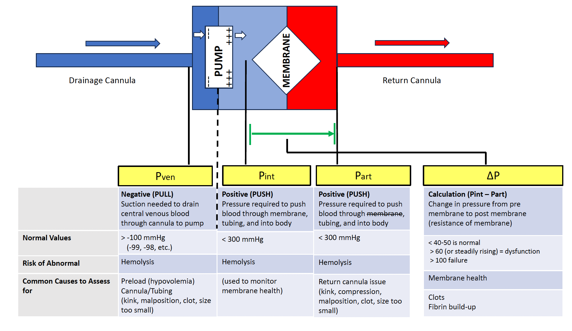
Themes for Weaning
VV ECMO
As lung function improves:
- Decrease extracorporeal gas-exchange support → assess whether the native lungs can maintain adequate gas exchange.
- A common final assessment involves stopping or substantially reducing sweep gas (often called a sweep-off trial or maybe a sweep trial) while blood continues to flow through the ECMO circuit (do not recommend stopping flow).
Note: this is different than an SBT. You increase vent settings, can maintain deeper sedation if necessary, and then simply turn off the sweep. - The ABG you are given would now represent how the patient is doing on the ventilator as the ECMO Console has been mostly "turned off" (flow is only to prevent coagulation at this point). Be sure you recommend "full" lung-protective vent settings, not "ultra-lung protective" settings.
VA ECMO
The question is different:
- Can the heart provide adequate circulation again?
- VA weaning focuses heavily on recovery of native cardiac function and hemodynamic stability as ECMO support is reduced.

