| 1. Basic Info |
- Caused by a virus thought to be Droplet Transmission but also uncertainty as to whether it is Airborne. (JAMA, WHO)
- While there are some predictable elements of COVID (such as ARDS in severe cases), variants and individual variability (including comorbidities, vaccination status, etc.) make it difficult to have a dependable list of symptoms, timelines of presentation, etc. In general, major symptoms often appear 4-5 days after exposure, perhaps as few as 3 days (omicron variant)
- Current evidence is mixed on how much immunity a person has once recovered - the presence of antibodies (Liu et al)
- While there are predictable patterns there are individual variations in how COVID presents. Severity ranges from asymptomatic to life-threatening (~2-3% mortality) (Wu & McGoogan):
- Mild to Moderate (similar to mild pneumonia): 81%
- Severe (dyspnea, hypoxia, > 50% lung involvement): 14%
- Life-Threatening (respiratory failure, shock, MSOF): 5%
- Common ICU admissions: hypotension (need for vasopressors) and hypoxemic respiratory failure (need for ventilator-support)
- Mortality rates on a ventilator are difficult to pinpoint as data is usually incomplete, different strains exist, etc. - numbers range from 16% to 88% (MGH, Meng et. al, Weis et. al)
- Evidence suggests presence of thromboembolic aspects (Berlin et al, Creel-Bulos et al)
- Comorbidities, as expected, may worsen disease severity and increase mortality - (Baum et al, Richardson et al)
A large percentage of hospitalized patients have comorbidities (Wiersinga et al)
- Typically ARDS will appear about a week after symptoms start (Berlin et al, SCCM, Wang et al)
- While respiratory system seems to be primarily affected, there are known extrapulmonary manifestations (Gupta et al)
|
| 2. Labs and Other Diagnostics |
Key (Critical Care) Labs (notice that most lab findings aren't all that sensitive to COVID, though they may provide hints)
| Lab |
Notes
|
| CBC |
Normal WBC count or leukopenia, ↑ WBC may be ominous sign (ACCP)
↓ Lymphocytes (lymphocytopenia) (CDC, SCCM, Guan et al)
↑ Neutrophils (neutrophilia) may be associated with increasing severity (ACCP, CDC)
↓ Platelets have been correlated with higher mortality (Ruan et. al)
|
| Biochemistry |
↑ BUN, Creatinine (ACCP)
↑ AST, ALT, Total Bilirubin (ACCP)
↑ CRP tracks closely with disease severity (and mortality) (Ruan et. al, Young et. al, CDC)
↑ D-dimer may indicate severity of disease, increase in mortality (ACCP, CDC)
↑ LDH may indicate severity of disease (ACCP, CDC)
|
| Biomarkers |
Procalcitonin may be normal initially, but severe illness may cause a (moderate) ↑ (CDC)
Significant ↑ may be more indicative of secondary infection, often bacterial, not viral COVID (ACCP, Lippi et. al) and is sometimes used to determine use of antibiotics with lower respiratory tract infections (many sources: Fabre et. al, Pink et. al)
↑ Ferritin may indicate the so-called "cytokine storm" crash some patients experience, indicate severity (ACCP, CDC, PW, Ye et al)
↑ Troponin is an indicator of severity of disease, suggest myocardial injury due to inflammation or COVID-19 directly (Gupta et al)
|
| Coagulation |
Various markers of coagulopathy may be abnormal: elevations in fibrinogen and D-dimer levels, and mild prolongation of PT/aPTT (American Society of Hematology)
|
| General Comments |
Note that complications are common: acute liver injury (elevated LFTs, however, do not appear to contribute to mortality MGH), acute kidney injury, acute cardiac injury, thrombotic events, shock, and should all be monitored (WHO)
The following increases may be associated with increasing severity: WBC, Neutrophil, ESR, D-dimer, LDH, CK, Troponins, Creatinine (ACCP)
|
- Imaging -
- Evidence of diffuse alveolar damage, pulmonary thrombosis (MJA)
- Arguments for CXR if resources are limited, otherwise consider CT scan (Fleischner Society, CDC, Pan et. al )
- Number of segments involved tracks closely with disease severity, as expected
- Imaging may be mostly normal to begin with
- Increasing numbers, extent, and desnity of ground-glass opacities on CT Scan may indicate disease progression (Zu et al)
- Ultrasonography - must be thorough for adequate information, amount of consolidation tends to trend with disease severity
- Presence of B-lines (ACCP)
- Subpleural consolidations (ACCP)
- Alveolar consolidation with air bronchograms, when severe (ACCP)
- Pulmonary Function Testing
- Current Trends:
- Consider performing screening (day before + day-of) to verify asymptomatic
- Preference for negative test when possible - avoid PFTs if positive test
- Symptoms, especially fever, should result in cancelation of testing, notify MD
- For Testing: Adequate (thorough) infection control in between patients, consider spacing tests of 30-60 minutes
- Long COVID: consider the role of PFTs for patients with lingering symptoms following acute infection (NDD)
- See other resources (ATS, ERS)
- Sleep Studies
|
| 3. Respiratory Clinical Approach |
This is an overview of strategy after reviewing conversations, emails, discussion boards, current various hospital procedures, social media groups, etc. As you can tell the sources aren't exactly "evidence-base" but more of a common consensus, so use them simply as "food for thought:"
- Acceptable isn't always Normal. Most are treating at a slightly lower SpO2 than normal, allowing for permissive hypercapnia, permissive hypoxemia, etc.
- Assess carefully. Not all COVID patients present the same. Treat the individual. Expect oxygenation to be an issue, either due to perfusion or due to ARDS-like process. Be watchful for V/Q issues which are more complicated than just administering O2.
- Consider reasonable efforts to support noninvasively, intubate when reasonable noninvasive efforts are ineffective
- Escalate O2 from NC to Venti/NRB mask
- Consider Lung Expansion and Airway Clearance as indicated, self-directed when clinically appropriate
- Consider noninvasive support (HFNC, perhaps CPAP/NPPV) as a trial
- Consider self-proning
- Keep head of bed elevated if not proning, sit out of bed, and mobilize as able
- Consider a trial of inhaled pulmonary vasodilators
- Assess often. Be watching for telltale signs of sudden deterioration. Patients can appear to improve, then "crash" again.
- Intubate when Necessary. There is a decision to be made ... balancing between the risk of premature intubation against the risk of sudden respiratory arrest with a chaotic intubation (Berlin et al). Signs of moderate-to-severe ARDS are unlikely to be turned around noninvasively. Significant comorbidities are an indicator of the need to intubate sooner.
- Work of breathing is probably a stronger indicator of need to intubate than just evidence of hypoxemia
- Tachypnea, on its own, may not be a primary indication for intubation (it is an expected physiological response)
- Consider trending: is patient stable or improving (consider observation vs. intubating) or is pt declining over time (favor intubating)
- Severe refractory hypoxemia (consider in context of other symptoms)
- Ventilate Carefully. Ensure lung protection. Watch driving pressures, recruitability. Overall, this means following ARDS strategies:
- Strongly consider proning
- Use of ARDSnet strategies is most common due to likelihood of ARDS-like phenotypes admitting to critical care (maintain Pplat 30 or lower, low driving pressures, use low VT, higher/optimized PEEP)
- Determine recruitability (consider R/I ratio - see topic below)
- Monitor driving pressures, maintain under 15
- Consider lung recruitability in general
- Expect a lengthy vent course as the norm. Extubating too aggressively may simply result in a reintubation. Be sensitive to the patient's "peak of illness." Initially, most advice was against performing tracheostomies, particularly open trachs, but some hospitals are now reporting frequent tracheostomy procedures due to vent days.
|
4. Clinical
Progression |
Inconsistency in Presentation
One of the challenges related to treating COVID-19 disease is that it appears to have a somewhat inconsistent presentation. Is it viral pneumonia? Is it ARDS? Is it a cytokine storm? Or is it a complex relationship of all those? In ICU there are reports of:
- Higher (not necessarily normal) vs. Lower compliances
- PEEP-responsive vs. PEEP-unresponsive
- Symptomatic vs. Non-Symptomatic Hypoxemia (Happy Hypoxemia)
- Thick, difficult secretions vs. Scant secretions
- Severe, rapid deterioration (cytokine storm?) vs. No deterioration phase, gradual improvement
Multiple Phenotypes Theory
Some have suggested multiple phenotypes for the virus (minimal data to support, some against), each with a different clinical progression. (Marini et al, Gattinoni et. al, ESICM)
Current Narrative: Gattinoni (theorized this, well respected out of Italy) even strongly warns against placing patients too strictly in one group over another.
Rebuttal: A study by Ziehr et al does NOT support phenotyping, but instead suggests a continuum consistent with ARDS (see MGH FLARE)
|
Phenotype L |
Phenotype H |
| Premise |
Hypoxia-driven dysregulation of pulmonary perfusion
(failure of hypoxic pulmonary vasoconstriction:
perfusion is redistributed to less-ventilated areas of the lungs) |
Pulmonary Edema/Collapse |
| Findings |
- Low Elastance (high compliance)
- Low V/Q
- Low Recruitability
- Low PEEP-response
- Low Lung Weight (primarily aerated)
|
- High Elastance
- Higher Recruitability
- High Right-to-Left Shunt
- Higher PEEP-response
- High Lung Weight (consolidation, equivalent to severe ARDS)
|
Potential
CT Findings |
well-aerated lung compartments |
lung aeration has decreased, much more ARDS-looking |
| Strategies |
Calculate Shunt such as A-a gradient
- Increase FIO2 (supplemental O2, etc.)
- Consider noninvasive (HFNC, CPAP, NPPV) as a trial
but be careful about pleural pressure swings (excessive inspriatory efforts)
- Consider intubation w/ sedation and possibly paralytics
- max PEEP 8-10 (higher may create deadspace, adversely redirect blood flow)
- "liberal" tidal volume (7-9 mL/kg) - some caution against (MGH)
- (Awake) Proning may help
- iNO may not be as helpful (esp if vasoplegic)
|
- Treat as severe ARDS with lung protection,
- higher PEEP but < 15
- Lower VT per ARDSnet (~5-7 mL/kg)
- Strongly consider Proning as a key intervention
- Consider ECMO, as indicated
- Consider trial of iNO
|
- In addition, patients may transition from Phenotype L to Phenotype H, theoretically due to the evolution of the underlying pneumonia-state as well as effects of "high-stress ventilation" including large pleural pressure swings. This transition is an important observation during care due to a possible change in strategy.
- Because there is this sense of "Happy Hypoxia" in some patients, many clinicians are considering hypoxia within the context of other symptoms (especially extensive inspiratory effort/WOB)
Cytokine Storming
This is an excessive immune response to the virus. In some patients, it is thought that cytokines and chemokines are rapidly increased, attracting inflammatory cells into lung tissue and causing brutal lung injury (Ye et al). Clinically this will present as an "aggressive" rapid deterioration (watch C-reactive protein and Ferritin) (Ruan). Note that cytokine storms have been associated with greater morbidity and mortality (Darden et al)
Thrombosis
- Coagulation dysfunction seems to be common
- Most deaths have some evidence of thrombotic DIC (look for dilated pulmonary vessels on CT; complaints of pleuritic pain) (MJA)
|
| 5. Personal Protective Equipment |
Remember: Protect yourself and others from potential exposures. Equipment must be worn correctly to be effective (See CDC 3 Keys for Respiratory Effectiveness, See CDC Mask Seal Self-Check)
Our Summary:
- Transmission occurs by infected respiratory secretions. This includes during exhalation (risk increases if increased exhalation such as with coughing, singing, sneezing, etc.). There is evidence that while the "6-foot" distance is reasonable, some exposure likely exists beyond that (CDC), and exposure over time (> 15 min) increases risks of such exposures.
- Studies of "viral load" support that greatest risk is closest to the patient, and when performing aerosol-generating-procedures. Patients more ill (ICU, for example) have a higher viral load and are more infectious.
Healthcare workers are often exposed to these more infectious patients.
- As always, infection control is only as good as the quality of the practice (appropriate size and technique, avoiding adjustments to PPE during patient care, removing equipment systematically, etc.)
Current Published Guidelines
We believe that clinicians at this point are aware of what PPE is needed when providing care for patients with COVID-19, but here are a few organizations providing guidance:
For aerosol-producing procedures, including much of what respiratory does, consider need-based care (more than usual), and then airborne precautions are recommended (including N-95 or above mask), as well as eye protection, gloves, gown, Negative Pressure room is ideal (CDC; WHO; SCCM). Some respiratory tasks that fall in this include:
- Manual resuscitation (bagging)
- Noninvasive ventilation
- Intubation/Tracheostomy
- Bronchoscopies
- CPR efforts (compressions + bagging)
- Airway clearance (including suctioning)
- Aerosolized drug delivery
- Proning a patient
- Sputum induction
|
|
6. Airway
Management
(Intubation, Management, Extubation)
|
Intubation
- Do not delay intubation if the patient is worsening or is pre-code (unstable). When to intubate is debated. (Ferioli et al, APSF)
- Intubation is high-risk for exposure
- There is no "Emergency" intubation - all PPE must be donned appropriately to minimize risk
Preparation
- Personal Protective Equipment: Airborne (a must!) + Contact = N95 or higher, gown, gloves, goggles/face shield. There is some evidence that intubation success increased with use of a PAPR (ASAHQ). Some recommend double-gloving primarily to use outer gloves to put laryngoscope blade in after intubation (APSF). Consider use of additional barriers (clear intubating box, usually plexiglass) when available/practical
- Least # people possible in room, close door during intubation and for a period after (AHA, WHO)
- Use of (preferably dedicated or disposable) fiberoptic equipment or whatever provides greatest chance of success on first attempt is highly recommended (ASA). It prevents exposure (looking directly into the airway) and improves chances of intubating successfully (see video demo from ATS) (APSF)
- Pre-oxygenating
- Team should have a discussed plan, all know part, closed-loop communication at all times. Consider Mock Intubations to practice (APSF)
- Assess patient for difficult airway - Upper Lip Bite Test has been suggested as most accurate (Cochrane)
- Because the goal is to minimize all exposures (and disconnections), the ventilator should be ready, in stand-by, resuscitation bag should be ready with filter
During Intubation
- Prioritize using the person who has the most experience/skill with intubating to avoid multiple attempts (ASAHQ).
- When possible, oxygenate for 5 minutes before RSI (using NRB if not bagging) (APSF)
- Expect no respiratory reserve (due to hypoxemic respiratory failure): SpO2 may drop quickly during attempt (Meng, et. al)
- Use Rapid Sequence Intubation (RSI) unless difficult airway identified (assess the airway if time) - RSI should decrease time, decrease cough-risk during attempt
- If possible, have 2 people confirm ET tube through vocal cords using video laryngoscope
- If unable to obtain airway, may need to provide GENTLE breaths with Resuscitation bag (if using), some recommend Max 2-3 attempts, then place supraglottic device (such as LMA) (Sorbello, et. al)
- Avoid disconnections - including administration of drugs via ET tube (AHA)
Post-Intubation
- Finger occlude ET tube as soon as it is placed and stylet out, then place on vent (or bag), Clamp ET tube if any delay (Kelly Clamps, 4x4 Gauze)
- Some hospitals report intubating, then placing directly on a ventilator for tube placement confirmation with ETCO2 (again, this avoids droplet aerosolization) = avoiding colorimetric capnography (EZ-Cap). If in-line ETCO2, not an option, consider using alternative methods of confirming placement - chest rise on vent, breath sounds, depth marking on ET tube, then follow-up CXR.
Extubation
- Most prefer a protected extubation which means treating as a very high-risk procedure and minimizing exposures at each step
- There are some anecdotal reports of patients having early relapses, so keep that in mind before extubating (GIVIT Mtg).
- Airborne and Contact precautions required - this is a high-risk for aerosolization procedure
- Limit the number in the room (preferably 2) (Tan, et. al)
- Recommended Procedure (Tan, et. al)
- Pre-oxygenate (FIO2 1.0 x 3 minutes)
- Consider using 2 "chux" pads, one on pt chest (pad side up), one over pt face (pad side down) - pad sides face each other.
- Gentle oral suction
- Suction through in-line suction via ET tube
- Remove ET tube stabilization/tie while holding ET tube in place
- Turn Ventilator to Stand-by or OFF (especially if it could auto-restart), cap end
- Deflate cuff and extubate - no further suctioning, do not instruct the patient to cough. Used ET tube should end up in between chux pads.
- Others (not source cited) recommend having in-line suction ON when deflating cuff, leaving suction on through the extubation maneuver to minimize aerosolization.
- Place O2 mask on patient immediately as tube is removed to minimize cough exposure (most recommend not using NC, consider NRB mask or Venturi mask)
- Consider using bag-mask over mouth/nose after extubation until coughing has subsided
- Consider transitioning to nasal cannula as patient settles and stops coughing
Tracheostomy
- Should trachs be performed? One Perspective (DeMaio et al, CHEST/AABIP/AIPPD Expert Panel Report)
- Recommended Resource: Global Tracheostomy Collaborative (COVID-19 Resources)
- Performing or assisting with Tracheotomies should be considered an aerosol-producing (highest risk) procedure with appropriate PPE, negative-pressure room
- Some report performing bedside (percutaneous) trachs
- Timing: consider no sooner than 2-3 weeks post-intubation, preference for negative COVID test (AAO-HNS)
- Consider use of paralytics (avoid coughing) (AAO-HNS)
- Consider use of Glycopyrrolate and lidocaine/epi to minimize secretions, bleeding (UMich)
- Percutaneous Procedure Elements (Niroula et al, UMich):
- Ensure FIO2 1.0 during procedure
- Care with dilatation (turn off vent, cover stoma between steps)
- When withdrawing, stop mechanical ventilation
- Withdraw ET Tube with guidance
- Inflate cuff, then restart ventilator
- Preference for cuffed (inflated), non-fenestrated tubes (AAO-HNS)
- Some recommend no dressing change unless evidence of infection (Pichi et al)
- Avoid trach tube changes when possible (AAO-HNS)
- Use of speaking valve requires cuff to be deflated if using (many avoid), consider surgical mask over nose/mouth (exhalation occurs through upper airway)
- Humidity: goal is to minimize aerosolization exposure, no real consensus on HME vs Heated Humidity for trachs (Austin)
Unplanned Extubation: Not an emergency - MUST take the time to put Airborne and Contact PPE on correctly. TURN OFF VENTILATOR as soon as safely possible. Place on NRB mask if not using manual resuscitation in hospital, if using consider 2-handed technique with bagging to avoid exposure. Assess patient for need for reintubation. (Tan, et. al)
Compassionate/Terminal Wean: Two main pathways we are currently aware of:
- Keep Intubated: CPAP +5, 21%
- Protected Extubation: Procedure as above, extubate directly to a mask (simple mask, for example), air vs. oxygen for comfort
Post-Mortem Extubation:
- If patient dies prior to extubation, consider airborne precautions (while no respiratory effort once vent is off, some are using utmost abundance of caution)
- Some recommend:
- Turn off ventilator
- Clamp endotracheal tube
- Disconnect the ventilator and immediately cap it
- Leave patient intubated, leave clamped, or consider immediately placing filter and cap on end of endotracheal tube
General Airway Management
All tasks should be performed with the goal of minimizing exposures, avoiding aerosolization of secretions
- Minimize disconnects from vent whenever possible
- When disconnecting from vent circuit:
- consider clamping endotracheal tube (use flat Kelly Clamp (no teeth with a 4x4 gauze) until reconnected to bag-valve-mask, etc. or cover the end of the vent circuit with a gloved hand while disconnecting to minimize aerosolizing droplets
- Some report a bolus of sedation if necessary (avoid coughing) (Sorbello, et. al)
- Some report placing ventilator in stand-by mode prior to disconnecting/clamping (Sorbello, et. al)
- If bagging at your facility, use a HEPA, viral/bacterial filter between Resuscitator Bag (BVM) and the artificial airway.
- If possible, avoid procedures that increase exposure-risk, including trach placement/change, bronchoscopies, etc.
- Ensure cuff pressures between 25-30 cmH2O (ensures appropriate seal with airway) - no evidence for over-inflating cuff
- Some reports of increased cuff leaks (causes are possibly due to higher PEEPs, proning)
|
|
7. Oxygenation Goals |
Recommended SpO2 Goals (WHO, GOLD, SCCM)
|
Adults with severe COVID
(distress, hypoxemia, shock)
|
Evidence Varies, with lower limits as low as 88% reported (Wax & Christian)
92-96% SCCM (SCCM suggests low threshold of 92%, but recommends that threshold be no lower than 90%)
90-96% WHO, UptoDate
88-94% AHA
|
| once stable |
> 90% |
| once stable, pregnant |
92-96% |
|
Pediatrics initially
(severe distress, cyanosis)
|
≥ 94% |
| once stable |
≥ 90% |
| Adults with COPD |
DO NOT WITHHOLD OXYGEN
Goal per normal (around 88-94%)
|
Upper limit is to avoid risks of hyperoxygenation (Cochrane)
- Interface: use of nasal cannula or mask interfaces seem acceptable, but avoid humidifying when possible (Ferioli et al)
- Consider having patient wear a surgical mask over the O2 delivery device to further minimize aerosolization (Ferioli et al)
- Encouraging patients to deep breathe, be out of bed sitting when possible, or mobilizing when appropriate may help (Thevarajan et al)
- Dyspnea may not be noted until PaO2 < 60, though tachypnea is a normal physiological response. Consider dyspnea a stronger sign of trouble (Tobin)
|
8. Noninvasive
Ventilation |
- There is some disagreement over the effectiveness and aerosolization risks around noninvasive in general
- When using, it is recommended that airborne PPE be used, consideration for negative-pressure or HEPA room
- Of noninvasive options, current evidence and practice supports HFNC over CPAP or NPPV
- No matter what, patient must be very closely monitored, preference for proactive intubation vs. emergent need for intubation (do not delay a needed intubation)
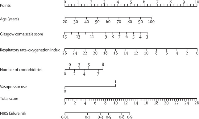
Potential Nomogram for Predicting Failure (Liu et al)
Patient prognostic values are located on the axis of each variable; a line is then drawn upwards at a 90° angle to determine the number of points for that particular variable. The sum of these numbers is located on the total score axis, and a line is drawn at a 90° angle downward to the NIRS failure risk axis to determine the likelihood of failure of non-invasive respiratory therapies. Alternatively, failure risk can be ascertained from the online calculator. Vasopressor use was represented on the axis at an arbitrary value of 1 (no use=0). NIRS=non-invasive respiratory support.
Considerations
- There is evidence that the risk is similar to normal supplemental O2 administration (Iwashyna et al, Leonard et al, Ferioli et al)
- General consensus seems to favor HFNC, or NPPV if HFNC not available ... when no indications to intubate (NIH)
- Must monitor closely: if significant indication of failure or instability, especially WOB: discontinue and intubate (NIH, Gattioni et. al)
- GCS < 10
- Increased vasopressor support
- P/F < 200 (also: Wang et. al)
- O2 should improve (not stay the same!) within about an hour
- Some recommend postponing intubation as long as possible due to poor coutcomes assoc. with it (See EVMS), but others still recommend early intubation (NIH)
- Others recommend avoiding noninvasive with ARDS due to lack of quality evidence, difficulty in maintaining low VT, difficulty in setting and tolerating "PEEP," risks of mucous plugging, etc. (MGH FLARE, Berlin et al)
- Use dual-limb circuits when possible (one limb is inspiratory, one expiratory)
- Mask interfaces should not contain a leak port (can't filter these, increase aerosolization). Instead, use either a dual-limb circuit, or consider using single-limb with a leak port - filter the port. Either way, an expiratory limb or "expiratory/leak port" is critical for patient to exhale.
- Helmet Interface
- See image: helmet interface - transparent helmet placed over patient's head, collar fitted carefully at base of neck. Held in place with straps at the underarms/axilla.
- May decrease exposure risk, may reduce intubation/mortality (Patel, Liu et al).
- Hazards include:
- Pressure/Noise to ears - consider earplugs with longer duration, higher pressures (Rodriguez et al)
- Rebreathing of CO2 - may result in higher CO2 retention, consider with hypoxic resp fail, caution if hypercapnia already (Rodriguez et al)
Note: anti suffocation valves (allow AIR in during interruption in flow) may decrease this, but also dips in oxygenation/PEEP
- Triggering issues/Autocycling in part due to helmet compliance - maybe better suited with CPAP since no trigger needed (Calvo et al)
Inspiratory effort may be exaggerated with higher WOB with helmet (Racca)
- Consider only with care as there's not a lot of data yet to support, some concerns in general (MGH FLARE)
- Interesting site that gives tons of info on this interface: helmetbasedventilation.com
High-Flow Nasal Cannula (HFNC)
- Primarily for acute hypoxemic respiratory failure associated with COVID-19 (pneumonia-like presentation)
- Benefits might include higher flows than other supplemental O2 (washout of dead meets patient demand, applies positive pressure to upper aiways), heat and humidity (assist with mucus hydration/clearance), resulting in less need to intubate although questionable impact on overall outcomes (Demoule et al, Ospina-Tascon et al, SCCM).
- Perhaps as effective as Helmet NIV (Grieco et al), may be more effective than standard O2 therapy but no more effective than CPAP (Cochrane Review)
- SCCM recommends (if supplemental O2 not working, prefers over NPPV), WHO recommends, AHA recommends at lower flowrates over CPAP/NPPV,
- Cochrane Review is inconclusive (insufficient evidence to support)
- Infection: Initially there were reasonable concerns about aerosolization risk, but most recent evidence supports HFNC being similar to other supplemental O2 delivery (See Crimi et al for review of evidence). This should be considered an aerosol-generating procedure (airborne precautions when possible) (WHO)
- Potential Modifications:
- Initial: Start at FIO2 100%, Flows of 50-60 L/min are commonly required (Crimi et al)
Noninvasive CPAP
- Increased mean airway pressure - while not using ventilators, doesn't augment breaths (supports lung protection)
- Achieving an adequate seal on mask can be a challenge, any leak increases risk of aerosolization/risk to healthcare providers, higher CPAP level may increase risk of leak, CPAP assumes minimal WOB - pH acceptable (as a reflection of PaCO2)
- Evidence: See this excellent article on CPAP for COVID (Josh Farkas).
- Modifications:
- Extra care with mask-fitting and adjustments (high-risk)
- If possible, avoid using a mask with an exhalation port, consider dual limb circuit (requires critical care vent which may use critical resources)
- Titrate CPAP to SpO2 goals - but do not exceed 20 cmH2O (gastric insufflation) - also remember, the higher the pressures, the greater the risk of leaks, and the tighter the mask the greater risk for skin breakdown issues
- Stop flow before removing device (Tan et. al)
Noninvasive Bilevel (BiPAP or NPPV)
- Poor mask fit, leaks around mask, taking mask off - all common and increase risk
- Mixed results, particularly with low P/F - anecdotal reports of success in New York, China, Italy
- High respiratory drive: self-injurious breathing. This occurs when rapid number of breaths, large breaths, with large pleural pressure swings can accelerate ARDS pathways (MGH)
- Evidence: NIH recommends if HFNC not available and no urgent indication of need for intubation.
- Modifications/Strategies:
- Dual limb preferred with filter on expiratory side
- If single-limb circuit, consider HME between exhalation port and mask (CTS) SEE IMAGE of SETUP
- Avoid using a mask with an exhalation port (single-limb use circuit with a leak port - filter it; dual limb - filter expiratory side)
- Maintain ΔP (IPAP-EPAP) for tidal volume around 6 mL/kg (SCCM, Vimeo - Dr. Clum) to avoid large transpulmonary pressure swings
- Stop flow before removing device (Tan et. al
|
9. Invasive
Ventilation |
Use an approach of treating what you see with an emphasis on lung protection. Avoid beating up on healthier lungs (higher compliance, nearly normal CXR) which may be more of a perfusion issue than a lung issue. We should avoid strictly protocolized care, preferring more of an individualized approach.
See AHA/AARC/ASA Algorithms for Basic and Advanced Ventilator Management
Strategies
There is some debate on appropriate vent strategies with the following all coming from reputable references:
- Adapting NIV Equipment for Invasive Ventilation (Dean Hess)
- Video: adapting NIV (not basic CPAP machines - which provide no ventilation support, difficult to provide higher FIO2s) for Invasive Ventilation (AARC)
- If single-limb NIV, ensure leak port is in-circuit, not mask, and filter it.
- Humidity is necessary - preferred is HMEF (HME + Filter together) - additional filters, etc. may increase mechanical deadspace
- Note that VT and minute ventilation displayed on single-limb is inspiratory, not expiratory (we can't measure expiratory at a leak port).
- Initial Settings (Hess at 6:15 mark)
- IPAP 25
- EPAP 12 (Note: driving pressure IPAP - EPAP is 13, adjust IPAP for 6 mL/kg IBW)
- Mode: S/T or PC
- Rate: 25/min
- TI: 0.8 s
- FIO2: 1.0
- The majority of resources still recommend ARDSnet, lung-protective strategies with higher PEEP (assuming the patient is PEEP-responsive)
- There is evidence to support maintaining the driving pressure (Pplat - PEEP) to under 12-15 (avg around 11). Driving pressure is a reflection of the functional size of lung and may be a predictor of mortality - above 15 is worrisome. Maintaining a lower driving pressure may help by minimizing over-distention and other complications. The bedside take-home? We should optimize PEEP and then titrate/drop our VT not necessarily to a mL/kg but by monitoring driving pressures (Amato et. al; Loring et. al, Brower)
TO CALCULATE: obtain Plateau Pressure, then subtract PEEP.
- Most reported prolonged periods of mechanical ventilation being required with extubation coming 8+ days after vent initiation (NEJM)
Equipment
- Specific Ventilators/Guidance:
- Equipment should be as closed-circuit as possible, HEPA or viral filters placed on any exhalation ports, preferably also inspiratory port (an abundance of caution). Dual-limb circuits are preferred over single-limb. Some hospitals have come up with scavenger-system add-ons to minimize exposures. Avoid disconnecting pt from the ventilator due to loss of PEEP, infection risk (WHO)
- Cleaning: With the use of appropriate filters, internal components should be safe to use between patients. Best practice would be to dedicate ventilators to COVID patient units. High-level disinfection should occur of all external surfaces. Internal sterilization is possible but usually requires quarantining the equipment and a labor-intensive process which is viewed by some as excessive processing (ASAHQ)
Quick Reference Vent Settings
Use this as a Starting Point. Note that most consensus statements recommend ARDSnet (AARC)
More explanation and details are available below the table
Recent data (MGH/Beth Isreal) does NOT support phenotyping, but instead a continuum leading to ARDS
|
Type "L"
Lungs More Normal (CXR, Compliance)
|
Type "H"
Lungs More ARDS-Like (CXR, Compliance)
|
| Mode |
No evidence to support any one mode (Cochrane)
Consider use of A/C due to heavy sedation (AARC)
Special Note on APRV: For those using APRV, here is a great resource on APRV-TCAV Method
|
| Tidal Volume |
More normal (~8 mL/kg IBW)
Note: Some advise against using
a higher VT (> 6) (MGH)
|
6 mL/kg IBW (IPC, WHO)
4-8 mL/kg IBW (NIH, SCCM)
|
| Rate |
Start under 20/min |
Around 20/min to start
Don't exceed 35/min
TI: ensure adequate exhalation, likely need lower TI
|
| PEEP |
Lower (< 10)
to avoid hemodynamic effects |
> 10
If PEEP-responsive, use optimal PEEP
Keep Driving Pressure < 15
Consider use of R/I Index
|
| FIO2 |
May need higher FIO2 |
Prioritize PEEP to wean FIO2, when possible
Higher FIO2s still likely needed for many
|
Tidal Volume
Note: This applies primarily to patients with presentations consistent with ARDS. Consider use of a lower PEEP, "normal" VT (8 mL/kg IBW), lower RR (< 20) strategy with healthier lungs.
Start at around 6 mL/kg Ideal Body Weight (IBW) (WHO, IPC). 4-8 mL/kg (NIH, SCCM)
If Compliance is normal, consider starting at 8 mL/kg IBW (ESICM)
- If Plateau is 30 or greater, drop tidal volume by 1 mL/kg IBW at a time until Pplat under 30 or at 4 mL/kg IBW (ARDSnet)
(do not go under 4 mL/kg IBW as this will approach dead space ventilation)
- As you decrease VT, you will likely need to increase RR. Consider increasing 5/min for each 1 mL/kg drop in tidal volume. For the most part we don't exceed a RR of 30-35/min due to air-trapping (not enough time to exhale)
- May need to actually INCREASE VT up to 8 mL/kg IBW (ARDSnet usually starts here) only if significant dyssynchrony or pH < 7.15 (WHO)
- Deep Sedation may also be required
Rate
Preferably start at whatever rate needed to match baseline MINUTE VENTILATION, but if initiating consider around 20/min to start, then titrate per ABG (see ABGs, below. See Inspiratory Time)
Avoid rates above 35/min (remember, do not normalize pH)
Inspiratory Time
Typically around 0.9-1.0 second, may need to consider shorter TI especially at 6 mL/kg IBW and below (0.7 or 0.6 sec).
Either way, ensure adequate exhalation (flow scalar should return to baseline) unless APRV where "therapeutic air-trapping" is employed.
PEEP
Use higher PEEP strategies if PEEP responsive (oxygenation improves with increase in PEEP level) (NIH, IPC, SCCM, WHO), despite some debate on whether it improves mortality (Cochrane). Use low PEEP ARDSnet table (AARC). Please see notes on phenotypes in disease section above. PEEP in non-ARDS may transmit to pleural pressures and have an exponential impact on (decreasing) cardiac output. There is evidence to support use of PEEP in patients with poor lung compliance, but the opposite is true if patients have near-normal compliance (normal plateau pressure)
- Higher PEEP is suggested to be > 10 (SCCM). If PEEP is too high: P/V Loop with beaking; cardiac output will decrease; BP will drop - this indicates overdistension of alveoli, DECREASE PEEP
- Perform an optimal PEEP maneuver if knowledgeable in how to do so
*Evidence for maintaining a driving pressure (Pplat-PEEP) of under 12-15 by adjusting VT and PEEP) if no spontaneous breaths (Meng et. al)
Evidence for use of a Recruitment/Inflation (RI) Ratio
Calculator
FIO2
Prioritize use of PEEP over FIO2 when possible. Increasing FIO2 without adequately recruiting alveoli (PEEP) will result in only minimal increases in PaO2. High FIO2s (1.0 or 100%) may result in further atelectasis from nitrogen washout
Sedation
There are reports that patients with COVID are requiring extra sedation. This may be in part due to ARDS-like processes, including lung-protective ventilator strategies. Metabolic processes related to critical care, longer ventilator course, etc., may also contribute to this need. Keep in mind that this may have an impact on weaning trials, time to extubation, delirium, and cognitive ability (see MGH FLARE on COVID sedation)
APRV
- Visit APRVnetwork for instruction and COVID-related guidelines (requires registration)
- Read MGH "FLARE" on use of APRV or HFOV (Advise against use of either APRV or HFOV) *this FLARE assumes spont breathing, see below
- TCAV Rescue Method (Quick Overview- read more at APRVnetwork above, if desired)
- PHigh: set to Pplat (from VC) or PIP (from PC) or 20-30 (starting on APRV, higher PHigh for more severe ARDS)
- PLow: 0 (if T Low is set to ensure therapeutic air-trapping, this will not result in derecruitment)
- THigh = ([60/current rate] - TLow) **use 1-4 if starting on APRV (lower THigh for more severe ARDS)
- TLow = 75% of expiratory flow (0.4 to 0.6 sec) or 0.2 to 0.6 if starting on APRV (lower TLow for more severe ARDS)
- Maintain PHigh for at least 24 hrs after FIO2 is weaned to 40%
- Increase THigh to maintain baseline PaCO2
- *May consider with heavy sedation with no spontaneous breathing (to avoid "self-injurious breathing" from high transpulmonary pressure swings. This may negate some of the MGH concerns in the linked FLARE.
ECMO
- The following is summarized from Bartlett et al:
- Consider when, despite interventions like proning, NMBA, and when there are no contraindications:
- P/F < 80 for more than 6 hours
- P/F < 50 for more than 3 hours
- pH < 7.25 with PaCO2 60 or higher for more than 6 hours
- Most appropriate for younger patients with minor or no co-morbidities
- E-CPR should only be attempted in places with experience, and then consider carefully
- There is general agreement with use as a rescue therapy after more conventional methods have been trialed (MGH FLARE) though some stay neutral on recommendation (NIH)
Humidity with INVASIVE Mechanical Ventilation
There is no consensus on Active vs. Passive Humidity (AARC). After reviewing articles and consensus, we have concluded that facilities are pretty evenly split between the two, with maybe an edge given to those using active humidity. There is a balance between what is best for patient (secretions, mucociliary clearance) and aersolization risk (disconnects of circuit). HMEs and HMEFs may be a bit drier and allow for less aerosolization but requires regular circuit-breaks with changing of HME, while active humidity may allow for less disconnect but increases risk of aerosolization (within a presumably closed circuit?). Equipment is also a consideration (some hospitals just don't have enough active humidifier setups).
Outside of COVID-19, we are not aware of any strong evidence for HME vs Active Humidity, most are split in terms of airway occlusion, mortality, incidence of secondary pneumonia (see a meta-analysis by Vargas et al)
- Passive Humidity (Heat and Moisture Exchanger, or HME):
- May result in more mucous plugging (be aware of need to instill saline and in-line suction, though instill may increase need to break circuit to replace HME)
- Consider vs. heated-wire particularly with single-limb ventilators (Zhonghua, et. al)
- Note that some HMEs may claim to be viral filters as well (HMEFs), but most do not (HMEs) - HMEFs may further protect vent/aerosolization
- When changing, consider pausing ventilator manually (do not use suction support with servo, for example which may autostart vent when disconnected), and clamp ET Tube. (HHS)
- Anytime HME is becoming occluded frequently with secretions, signs of inspissated secretions, etc., strong consideration for active humidity instead (AARC)
- HME may interfere with flow in APRV
- In cases of increased airway resistance, secretions are thick and hard to manage, higher minute ventilations, HME may be ineffective
- HMEFs increase deadspace by about 30 mL, with recommendation for adjustment in set VT in moderate-to-severe ARDS (AARC)
- Active Humidity (such as a heated passover humidifier):
- May decrease the number of vent circuit disconnects vs. HMEs
- May result in more condensate in tubing, AVOID breaking circuit to empty condensate (exposure risk), used closed-system trap if available. Adjust humidity if able, consider room temp, etc.
- May increase risk of aerosolization (heat + humidity), though some suggest it creates water vapor which can't transmit COVID-19 (Fisher & Paykel)
- Many facilities don't have enough active humidifiers for every ventilator, so it may make sense to have a transition plan from HME, this will be high risk changing between the two (anytime the circuit is broken)
- May be more appropriate wiith mucociliary transport, management of secretions, prevention of VAP (see white paper from Fisher & Paykel)
- Some recommend active humidity for patients on ventilators for longer periods (so most COVID patients)
|
|
10. Weaning/
Discontinuation
Extubation Procedure is covered in above section on Airway Management
|
See: Our Summarized Algorithm for Weaning
- Reports are that some wean off around 8-10 days, but that many require 15-20 days of ventilation before being ready to discontinue from vent (NEJM, MGH) - patience.
- Once P/F ratio improves and ventilator settings, especially FIO2 and PEEP have been normalized, consider weaning. (MGH FLARE)
- Once patient meets checklist criteria to wean - consider protocolized weaning, coordinated with sedation vacation (MGH FLARE, PAHO)
- Consider use of PSV to wean, evaluate carefully for tolerance. Remember, there is some concern about "self-injurious" breathing from large pleural pressure swings, so consider when to use PSV cautiously (has patient "peaked" in illness, for example?) (MGH FLARE, Brochard et al)
- Criteria to Consider
- Meeting oxygenation goals with FIO2 < 0.4, PEEP < 8-10 on PSV < 10 (Tan et. al)
- Secretions should be minimal/manageable (some reports are lots of secretions for COVID pts, others report minimal)
- Some recommend using less-aggressive weaning (goal to avoid need for reintubation), using higher "thresholds" to extubate (MGH, See also MGH FLARE)
- Ability to cough
- Mental status (open eyes, grasp hand, stick out tongue)
- Some recommend use of cuff leak test before extubating (about 10% have post-extubation stridor)
- Varying expert opinion/data on bridge therapies like HFNC (MGH FLARE, Zhu et al, Zhao et al)
- Avoid use of T-Piece trials, including with trach patients (as well as trach mask). Use pressure support weaning instead (CTS)
- Note that you may be able to wean some patients to minimal support (okay for extubation) but should consider their inflammatory state and whether the patient has "peaked" - consideration for not extubating until reasonably sure that period has passed (Emory)
- Slowly emerging thoughts on post-acute weaning (such as weaning units) (Levy et al)
- Should be trached before transfer
- Should be transitioned to appropriate ventilator model
- Should be stable (P/F > 200, no need for proning, no pressors, no major organ failure)
|
|
11. Critical Care
Strategies
(Proning, Recruitment, inhaled pulm vasodilators, ECMO)
|
PRONING
There is a strong consensus that proning a patient is a critical step in management, even pre-intubation
(Proning may improve ventilation-perfusion matching which can increase oxygenation)
Proning is considered an aerosol-generating procedure due to the risk of coughing/disconnect. Use Airborne PPE if possible
Self-Proning (also called Awake Proning, Conscious Proning)
- Consider having patients self-prone with abnormal V/Q - ARDS-like (Elharrar et al, Telias et al, Perez-Nieto et al, Scaravilli et al, Caputo et al)
- This is a supportive technique, not treatment, observe for signs of decline carefully (Caputo et al)
- Goal should be early application, helpful with mild-to-moderate ARDS, not recommended for severe (P/F < 100) (Ding et al)
- One article recommends early warning (start self-prone): RR>30, SpO2 < 93% RA, or HR > 120 (Sun et al)
- Contraindicated for severe distress or indications for need to intubate immediately, altered mental status, unstable spine, recent abdominal surgery, thoracic injury (NIH)
- See algorithm for use (ICS, pg 5)
- See basic video suggesting one way to instruct patient
- Should be performed with supplemental O2 in place or on high-flow nasal cannula. May work with CPAP/NPPV. (Sartini et al)
- Sessions of at least 30 minutes are recommended, max is as much as tolerated. Goal should be 1-2 hrs, then change position (ICS)
- One suggestion is 30-120 mins in each of the following, keep cycle (ICS):
- fully prone (bed flat)
- right-side (bed flat)
- sitting at 30-60 degrees (head of bed up)
- left side
- fully prone
- We have not found any reports of adverse effects, although intolerance is noted for some pts (Scaravilli et al)
- If pregnant or obese, modify position to lateral decubitis
- Instruct patients to sleep in lateral decubitis or prone position, avoid supine
- With any worsening in oxygenation in prone, discontinue
- Primary improvement is in oxygenation in as early as 5 mins (Caputo et al)
- Some evidence this decreases need to intubate - one study that it decreased intubation by > 60% (Ding et al, Scaravilli et al, Caputo et al)
Critical Care (Ventilator) Proning
- Emphasis is on early proning (Ding, et. al; Sun, et. al; Agrawal, et. al; Kallet, et. al)
- Some recommend proning if refractory hypoxemia (SpO2 < 90%, P/F < 150) despite maximizing vent settings (AARC, ATS)
- If pregnant or obese, use lateral decubitus position
- Deep Sedation (RASS -4 to -5) should be considered while proning
- Prone for no less than 6-hours at a time (multiple sessions per day), but preferably 12-20 hours/day (JAMA, NIH, WHO, SCCM)
- Some recommend discontinuing prone once patient is sustained in stable state (FIO2 needs, Pplat, ABG, P/F)
- The airway in Prone: Reports of lost airways (gravity, secretions), skin breakdown (facial edema). Consider:
- Secure with cloth tape - video demo of that here (use 2 people to tape, change when patient supine)
- Reinforce with "pink tape" (such as Hy-Tape), esp if issues with oral secretions
- Some also recommend successful use of "Stabilock tube holder" and "COMFIT"
- In one report, Hollister in prone was linked with breakdown requiring treatment (Gomaa and Branson)
- Use an "omniflex" adaptor, if needed
- No matter the method used (or device used), skin integrity should be monitored closely due to facial edema in prone (Gomaa and Branson), perhaps consider prophylactic dressings (Peko et al)
- CPR IN PRONE: If no airway, turn supine. If airway, CPR in prone (unless no risk of exposure in flipping to supine). SEE CPR TOPIC, BELOW
Proning Resources
Other Feedback/Information
- Airway: RT should always be present to prone or supine a patient - securing the airway and avoiding disconnect is absolutely critical
- Most people report performing the "swimming" method of proning - turn head side-to-side every 2 hrs (See Oliveira, et. al for photos/description of this)
- Lack of overall proof regarding proning (not a lot of evidence to support it), but thusfar evidence is promising (Cochrane)
Recruitment
The concept of "recruitable lungs" suggests that some lungs are "recruitable," others are less-so (also referred to as PEEP-responsiveness). Gathering evidence can be one step in determining this.
Recruitment/Inflation (R/I) Index
- A calculation that estimates the potential for lung recruitment (Chen et al, Tommaso et al)
- A single-breath maneuver that can be performed bedside
- Values > 0.5 may suggest the potential for lung recruitment
- Steps and Calculator
Recruitment Maneuvers
- Use with caution (watch closely for any desaturation, hypotension, barotrauma. STOP maneuver if any of these occur) but some recommend use of a recruitment maneuver. (SCCM), Cochrane review suggests no improvement in 28-day mortality.
- Suggested: Place patient on CPAP 35-40 cmH2O for 40 seconds, some recommend less time for safety - like 15 seconds (ESICM)
- Do NOT: Perform Incremental PEEP recruitment (increasing PEEP from 25 to 35 to 45, each level for 1-2 minutes) (SCCM)
iNO or inhaled prostaglandins
- Avoid routine use of inhaled vasodilators (use only when PEEP, recruitment, and proning are ineffective) (AARC)
- Aerosolized vasodilators should be avoided. (AARC)
Inhaled Nitric Oxide
- There has been growing clinical interest in the use of inhaled Nitric Oxide (iNO) with two possible application benefits:
- Antiviral: iNO showed antiviral properties against SARS, if effective may decrease severity, length of illness
- Pulmonary: treatment of pulmonary hypertension with improved hypoxia
- Some mixed recommendations, but a short trial with signs of severe ARDS may be indicated (NIH, SCCM, MGH)
- Anecdotally, RTs and MDs are reporting rapid improvement in at least some patients receiving iNO.
- Several studies are currently being performed: 1) CPAP or NRB with iNO, 2) Intubated, 3) Prevention of COVID in Healthcare Workers
Epoprostenol and Other Inhaled Pulmonary Vasodilators
- Really minimal evidence of improving overall mortality with ARDS (Cochrane)
- Some report using vasodilators through a setup like an Aerogen via a syringe pump, with expiratory filters
- Anecdotal reports of response rates are "sometimes" (maybe around 50%?)
- As with iNO, a short trial (around 2 hours) should be performed. If no effect in oxygenation (PaO2), discontinue
- Attempt to wean after 1-2 days, then daily
NMBA
to manage ventilation of COVID patients, some combination of deep sedation and possibly paralytics might be necessary, especially if:
- Dyssynchrony with ventilator
- Plateau Pressures over 30 despite other attempts (such as PEEP, low VT) to lower
If needed, consider boluses vs. continuous initially. If persistently high plateau pressures, consider 48-hours of continuous SCCM
ECMO
- Consider long-term (> 6 hours) ECMO for either respiratory failure or cardiopulmonary failure, especially when other options have all been trialed (FDA, ATS, WHO, SCCM), NIH stays neutral (no evidence for or against)
- As indicated, use is primarily for "rescue" not prophylactic, overall results are inconclusive (Cochrane)
- Use is considered "salvage" and should be performed before evidence of multiorgan failure (Gupta et al)
- Evidence is for early incorporation before lengthy vent course, avoid use in elderly (ELSO)
- Variation in reported outcomes, anywhere between 50-86% mortality despite ECMO (Gupta et al)
- See MGH ECMO Protocol
- See extensive article, "Planning and Provision of ECMO Services" (Ramanathan et al)
Multiple Patients on a Single Ventilator
Our official position is in line with the majority of respiratory and medical professionals: Multiple Patients should not be put on a single ventilator. The risks (including causing damage to all lungs being ventilated, increased cross-contamination, exposure risks due to more frequent disconnects) outweighs the benefits (stretching limited equipment/resources). MAJOR considerations include the need for both sets of lungs to have very similar lung mechanics (compliances, resistance, respiratory quotient, BMI, etc.), the driving pressure (Pplat-PEEP) needs to be identical, and the needs the same (acid-base, including metabolic component to a degree, PEEP, inspiratory time, total rate). If hypothetically able to match and maintain match (nearly impossible), these variables would need to stay the same in both patients the entire time (impossible). At best this would require a Respiratory Therapist be very close to bedside, constantly monitoring, all the time.
A major joint statement has been published on the matter:
See the Joint Statement on Multiple Patients per Ventilator (SCCM, AARC, ASA, ASPF, AACN, and CHEST)
See article: Personalized Ventilation to Multiple Pts Using a Single Ventilator (Han et al)
|
|
12. Troubleshooting
Please let us know if there are troubleshooting areas you are facing that aren't listed here yet.
|
| Signs/Symptoms |
Possible Causes |
Possible Strategies |
|
Secretions thick, tar-like
Difficulty weaning despite evidence of resolving illness
|
Reports of thick secretions coating ET tube |
- Consider tube exchange (aerosolization risk)
- Consider active heated humidity (aerosolization risk)
|
Low PaO2 or SpO2, otherwise "healthy" looking
(WOB mostly normal, Chest imaging mostly normal, etc.) |
"Silent or Happy Hypoxia" |
- Avoid injury to the lungs, use noninvasive as bridge
- Avoid intubating, if possible (read Tobin)
- Consider (awake) proning
|
|
Sudden Deterioration on Ventilator
|
- Reports of higher incidence of pneumothorax with SARS in general, but may be less with COVID - higher lung compliances (Wax et al)
- Reports of patients suddenly deteriorating as they reach so-called "peak" of illness.
- Potential asynchrony
- Deterioration after respiratory improvement may indicate cardiomyopathy
|
- Always check basics: airway placement, airway patent, bronchospasm, V/Q change with recent pt position change, etc.
- Suspect and rule out pneumothorax
- Suspect and rule out pulmonary embolus
- Verify synchrony on ventilator, some reports that sedation requirements can be high, delirium high
- Check and correct other indications of high O2 demand (such as fever)
- Check for metabolic influences (esp non-anion gap)
- Adhere to ARDSnet strategies if meeting definition of ARDS
- Strongly consider proning, if not already
- Watch for flow starvation, esp if neuro involvement (Worsham et al)
- Suspect and treat for co-infections, comorbidity involvement
|
| Difficulty in Weaning |
- Underlying illness hasn't "peaked"
- Respiratory muscle weakness
- ET Tube with thick, dried secretions (see above)
|
- May take extended period of time to wean/extubate/liberate
- Consider bedside trach (controversial)
|
Air-Trapping
(auto-PEEP) |
More common when bronchospasm or inadequate exhalation time (such as when set rate is high > 30), I:E ratio is inappropriate (should be 1:3 to 1:4, may need 1:5 with obstructive disorders like COPD or CF) |
Check flow scalar (does exp flow return to baseline?), I:E ratio (at least 1:3 to 1:4?), then decrease TI (or decrease set rate if TI already short)
Consider need for bronchodilator (wheezing, etc.)
Consider mode (if APRV/Bilevel, some air-trapping is considered therapeutic, exp flow termination should be about 75% of PEFR)
|
|
13. Airway
Clearance |
Bronchoscopy
High-Risk due to potential exposures: priority use of PPE (N-95, Face shield, gown, gloves) - avoid use if possible!
- Bronchoscopy in COVID+ is relatively contraindicated - use only if upper resp samples are negative and needed for significant clinical management (AABIP, IPC)
- Postpone all non-urgent bronchoscopies (AABIP, ACS)
- Sputum specimens should be obtained by closed suction with endotracheal tube with COVID sampling preference for lower respiratory tract (SCCM)
- If considering: minimize disconnections, use of bronchoscopy adapter on ET tube is recommended
- If considering: place mask on patient during bronch if not intubated (minimizes exposure)
- Consider suction catheter in patient's mouth to create a local negative pressure (Ferioli et al)
- Use of disposable bronchoscope, if possible
- Minimal personnel (only essential people needed to perform bronch)
- For all samples sent to lab: be sure to alert lab to COVID-19 status
- Standard high-level disinfection for equipment
Suctioning and Bronchial Hygiene
Use of airway clearance to address excessive secretions, difficult to clear secretions, may be necessary but consider infection control due to increased risks associated with aerosolization (WHO)
Strong preference for closed suctioning only (in-line suction) when intubated (WHO, CTS)
minimizes aerosolization exposure, decreases derecruitment of lungs (alveoli collapse)
Do not induce for sputum collection (such as use of hypertonic) (AABIP)
Avoid open suction techniques, including nasotracheal suctioning, open suction of the tracheostomy/stoma
If absolutely necessary, treat as a critical activity with priority on PPE (N-95 mask or higher level, face shield, gown/gloves).
Consider use of self-directed bronchial hygiene, whenever possible (Acapella, for example)
|
14. Aerosolized
Therapies |
Use MDI with spacer versus a nebulizer. One study showed active virus 3-hours in air sample after a nebulizer treatment (assuming not in a negative pressure room) (Khoo et al; Munster et. al)
- There is no role for inhaled bronchodilators with COVID-19 unless comorbidities (AARC)
- Aerosolizing drugs/treatments should be considered HIGH RISK *treat as Airborne with N-95 or higher mask (CMAJ, CP)
- Avoid combining aerosol therapy with airway clearance (Arzu)
Choosing a Drug-Delivery Therapy (in recommended order of preference, based on exposure risk, not drug deposition, etc.):
- Use an MDI (with spacer) when possible. Consider shared cannister protocol to preserve aerosolized drug availability. Use MDI adaptor/HME on vent circuit.
- Consider breath-actuated neb (BAN) with filter, or a breath-actuated vibrating-mesh neb to minimize number of breaths required (for example, if MDI unavailable)
Specific to: 2.5 mg (0.5 mL) of Albuterol, with no saline and no additional drugs
- Use a filtered nebulizer (such as those used with pentamidine, or with HEPA/viral filter placed), preference for mouth-piece over mask (Arzu)
- Consideration for surgical mask over interface if administering by HFNC (Arzu)
- On Vent, consider vibrating mesh neb in-line with extra filter at the expiratory port during treatment (CTS, Arzu)
|
|
15. Protected
Code Blue
|
Direct Link to the COVID-19 AHA new Algorithms (pg 14+)
Other specific recommendations: AHA
All CPR activities should be performed using Airborne Precautions
Protected Code Blue
- While initial steps to defibrillate may occur without all PPE, those providers should be replaced with providers with appropriate PPE as soon as possible. The patient may be masked if that can be done without delaying or interrupting compressions
- When rescue breathing is necessary, use of a HEPA filter with bag-mask ventilation is recommended (AHA)
- Attempt to minimize number of people in room
- Use mechanical CPR device if available, if pt meets height + weight criteria, to minimize personnel needed (AHA)
CPR with an artificial airway
- Consider leaving on ventilator during code to keep a closed circuit and make the following changes (per AHA guidelines):
The AHA is supporting a model of asynchronous ventilation - as evidence has been spotty about the benefits of breaths anyway (which is why "compression only" CPR is an approved method of layperson CPR) which has to be balanced with the very real threats of exposure to healthcare workers. In addition, Vent outcomes are poor with COVID, CPR outcomes are even poorer.
- Mode: PC, A/C with target of 6 mL/kg IBW (avoid alarms and high pressure within the circuit)
- FIO2: 1.0
- Trigger: OFF
- Set Rate: 10/min (adults, peds) or 30/min (neo)
- Consider PEEP to optimize for venous return to heart (maybe 8-10, discussed, not published)
- If/when ROSC is established, place on appropriate clinical settings
- Defibrillating on Ventilator
- Risk: while extremely rare, reports of fires/arcing in the presence of an oxygen-enriched environment
- Reason to Consider: any disconnect of the airway/vent circuit increases aerosolization risk to those in room
- Modifications to Consider if Leaving on Vent:
- Drop O2 below 50% and ensure any exhalation is 30 cm (12 in) away (APSF)
- Consider pausing the ventilator during defibrillation - extra measure of safety despite closed circuit (APSF)
- DO NOT disconnect the vent circuit and leave on patient - increases risk (AHA, ECRI)
CPR for Patients who are in Prone Position (AHA)
- No Airway: attempt to place in supine position for resuscitation
- Artificial Airway: avoid turning the patient to supine unless able to do so without risk of equipment disconnection
- Place defibrillator pads in the A-P position
- Provide CPR with pt remaining prone - over T7 and T10 vertebral bodies
|
|
16. Respiratory/
Critical Drugs
|
Corticosteroids
There has been much discussion around the use of steroids with COVID. There is no current evidence to support avoiding steroids, especially inhaled (patients with Asthma, COPD, etc., should continue to take steroids as prescribed/recommended).
Consider avoiding drugs that induce cough whenever possible (mucokinetics such as acetylcysteine, hypertonic salines)
Consider careful use with patients with abnormal secretions related to diseases - Cystic Fibrosis, Bronchiectasis, as needed when appropriate PPE is available, negative pressure rooms are preferred
If thick secretions, some recommend use of 3% saline (closed-circuit) or possibly guiafensin (by NG/OG)
Several studies are looking at iNO as as option (see critical care strategies section).
Asthma Exacerbation: Consider use of Albuterol by MDI with spacer initially for exacerbation, every 20 mins x 3 doses. Consider IV mag if needed. Avoid continuous albuterol due to aerosolization risk unless absolutely necessary. Be aware of need for early intubation (CHOP). Systemic steroids should still be considered in severe asthma exacerbations (benefits may outweigh risks) (WHO)
|
| 17. Transport Considerations |
- Consider early transfer of deteriorating patients to ICU (Liew, et al.)
- Medically-essential transport is preferred
- Inform receiving department of COVID+ patient beforehand
- Transporting Team should wear PPE (some recommend all everything (Liew, et al.), CDC recommends full PPE while handling the patient, but then only wearing a face mask during transport
- Patient should wear surgical mask if not intubated or be in a portable tent system (no mask needed in that case)
- Clean and disinfect all high-touch surfaces, including side rails, headboard, footboard, etc. (NebraskaMed)
|
| 18. Home Care |
CPAP and NPPV considerations
- Consider hands-off approach
- Videos and other resources to learn about the equipment initially (interactive training modules are ideal)
- Telehealth-style consultation/call to ensure appropriate use and understanding (where billing allows)
- Equipment then picked up, preferably as a curbside at home care company, or delivered to patient's front door.
- If pt is COVID+, they should use CPAP at home, but sleep in a separate bedroom, preference for separate bathroom (AASM, Baker et al)
- Change filters as normal
- Instruct in diligent hand hygiene practices
- Keep cleaning and disinfecting mask and hose (Philips, ResMed)
Use of Home Equipment in Hospital/LTAC (particularly for OSA)
- Consider transition to hospital equipment per guidelines in noninvasive above (for filtering, use of non-vented mask, dual-limb circuit, neg. pressure, etc.)
- Some not allowing use of home CPAP equipment with COVID patients due to exposure risks
|
| 19. Specific Comorbidities |
Obesity
Higher mortality. Aim for prevention when possible (self-prone, mobility) (ASA)
|